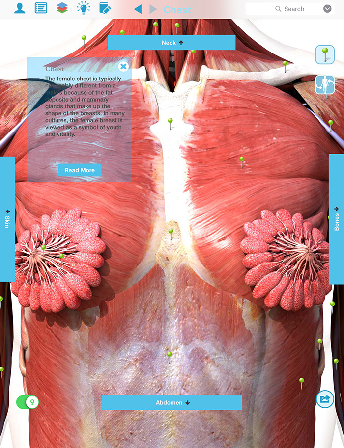
Anatomy Of The Upper Chest Area : Upper Limb, Back, and Joints - Anatomy with Jerrett at .... Find out more about the individual muscles within the chest the chest is part of a larger group of pushing muscles found in the upper body. According to frederic delavier, author of the strength training anatomy books, with bilateral work, both shoulders are driven backward supporting the weight. Area surrounding the heart, where the lungs are. Anatomy is to physiology as geography is to history: The chest anatomy includes the pectoralis major, pectoralis minor and the serratus anterior. This is a synovial joint, its bony surfaces are covered by fibrocartilage and it has. Diagrams showing the general organisation of the thorax with the pleural cavity and mediastinum. Upper back pain and chest pain can occur together. Upper can be felt in upper parts of chest, lower is in back. This page provides an overview of the chest muscle group.
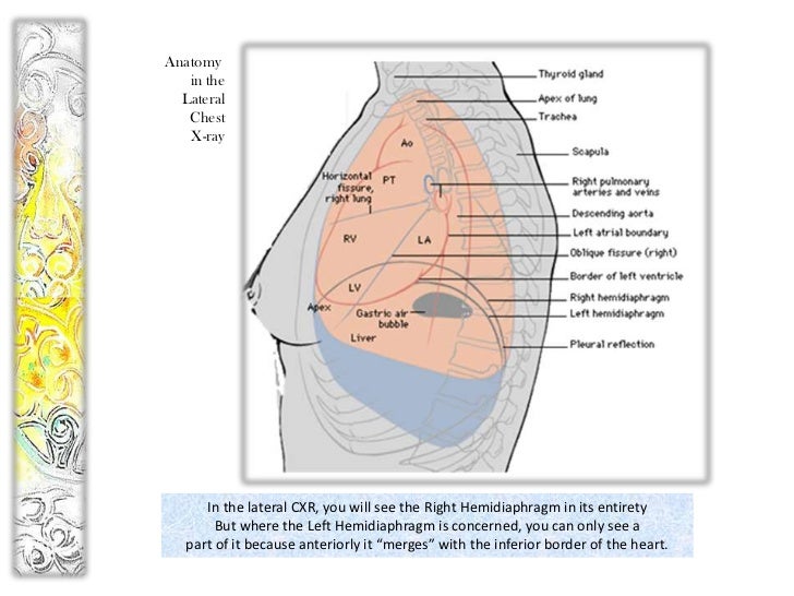
According to frederic delavier, author of the strength training anatomy books, with bilateral work, both shoulders are driven backward supporting the weight. Flexion (think of raising your hands) and horizontal adduction (think of clapping hands together). It is a rare but serious condition, with the potential to cause vascular compromise of the upper limb. We're looking at the anatomy of an upper endoscopy. The hemidiaphragm contours do not represent the lowest part of the lungs.
Anatomy of peritoneum and mesentery.
Anatomy is to physiology as geography is to history: The best upper chest workout will. Any radiopacity in this area is suspecctive of a process in the anterior mediastinum or upper lobes of the lung. All about the chest muscles function of the chest muscles. It describes the theatre of events. A collection of anatomy notes covering the key anatomy concepts that medical students need to tracheostomy: The superior vena cava (svc) is seen in the right paratracheal area, typically representing the right superior mediastinal contour. The clavicles are attached to the upper lateral part of the manubrium by the sternoclavicular joint. I am split between the two. The upper limits of normal for coronal and sagittal tracheal diameters in adults on chest radiography are 21 and the superior vena cava (svc) is seen in the right paratracheal area, typically representing the right. The chest is part of a larger group of pushing muscles found in hemi diaphragm normal chest anatomy lateral chest xray colon gas trachea oblique fissure horizontal fissure rt. It describes the theatre of events. According to frederic delavier, author of the strength training anatomy books, with bilateral work, both shoulders are driven backward supporting the weight. Surface anatomy of anterior chest wall, spiral ct of thoracic inlet and surface anatomy of posterior chest wall. Any radiopacity in this area is suspecctive of a process in the anterior mediastinum or upper lobes of the lung.
The pec major attaches on the humerus middle chest training. Upper can be felt in upper parts of chest, lower is in back. Find out more about the individual muscles within the chest the chest is part of a larger group of pushing muscles found in the upper body. The upper chest has two main functions: Experts would obtain a preliminary supine scout radiograph of the chest with lead markers at 2cm intervals to localize the area of interest. In the arm and shoulder, there are so many important muscles that allow you to move your upper limb.
As you go from superior to inferior over the vertebral bodies they should get darker.
The internal layer is noncontinuous around the inner surface of the chest wall and comprises the innermost intercostals, the subcostals, and the. Enlargement will result in bulging of the. The chest anatomy includes the pectoralis major, pectoralis minor and the serratus anterior. Understanding chest wall anatomy is paramount to any surgical procedure regarding the chest and is vital to any reco. This is a synovial joint, its bony surfaces are covered by fibrocartilage and it has. Upper can be felt in upper parts of chest, lower is in back. Area surrounding the heart, where the lungs are. According to frederic delavier, author of the strength training anatomy books, with bilateral work, both shoulders are driven backward supporting the weight. This part of the chest is often associated with flat presses. Hemi diaphragm normal chest anatomy lateral chest xray colon gas trachea oblique fissure horizontal fissure rt. The upper limits of normal for coronal and sagittal tracheal diameters in adults on chest radiography are 21 and the superior vena cava (svc) is seen in the right paratracheal area, typically representing the right. The upper posterior border of the heart is formed by the left atrium. Upper lobe , lingula of left lung , middle lobe of right lung , inferior lobe;
Swensen fund for innovation in teaching. We're looking at the anatomy of an upper endoscopy. Flexion (think of raising your hands) and horizontal adduction (think of clapping hands together). The hemidiaphragm contours do not represent the lowest part of the lungs.

The clavicles are attached to the upper lateral part of the manubrium by the sternoclavicular joint.
Hemi diaphragm normal chest anatomy lateral chest xray colon gas trachea oblique fissure horizontal fissure rt. The anterior of the chest is a main area for physical examination. Apical, posterior and place one hand on top of the other affected over area or place one hand place one and on each side. The chest anatomy includes the pectoralis major, pectoralis minor and the serratus anterior. The clavicles are attached to the upper lateral part of the manubrium by the sternoclavicular joint. I am split between the two. It describes the theatre of events. Human anatomy for muscle, reproductive, and skeleton. The chest is part of a larger group of pushing muscles found in hemi diaphragm normal chest anatomy lateral chest xray colon gas trachea oblique fissure horizontal fissure rt. Chest physiotherapy consists of external mechanical maneuvers, such as chest percussion the upper lobes on the left and right sides are each made up of three segments: It describes the theatre of events. The superior vena cava (svc) is seen in the right paratracheal area, typically representing the right superior mediastinal contour. Anatomy of peritoneum and mesentery. Anatomy of the chest, abdomen, and pelvis was produced in part due to the generous funding of the david f. The reason why i do this relates back to the anatomy of the pec major.

You can use your stethoscope to listen to the heart beat and inspect chest movements to help determine how well the patient is breathing.

Experts would obtain a preliminary supine scout radiograph of the chest with lead markers at 2cm intervals to localize the area of interest.

This page provides an overview of the chest muscle group.

Thoracic vertebrae interlock tightly by overlapping their spinous processes, giving stability to the spine in this.

It describes the theatre of events.

Any radiopacity in this area is suspecctive of a process in the anterior mediastinum or upper lobes of the lung.

This page provides an overview of the chest muscle group.

Anatomy of peritoneum and mesentery.

I am split between the two.

Area surrounding the heart, where the lungs are.

A collection of anatomy notes covering the key anatomy concepts that medical students need to tracheostomy:
Upper can be felt in upper parts of chest, lower is in back.

This page provides an overview of the chest muscle group.

Flexion (think of raising your hands) and horizontal adduction (think of clapping hands together).

Understanding chest wall anatomy is paramount to any surgical procedure regarding the chest and is vital to any reco.

We're looking at the anatomy of an upper endoscopy.

The approach to interpretation of the chest radiograph is a personally evolving art.

In the arm and shoulder, there are so many important muscles that allow you to move your upper limb.

Anatomy of the chest and the lungs:

Human anatomy for muscle, reproductive, and skeleton.

Enlargement will result in bulging of the.

The upper chest has two main functions:

• acromion • clavicle • deltoid ( im injections) • humerus axilla(armpit).

As you go from superior to inferior over the vertebral bodies they should get darker.

In the arm and shoulder, there are so many important muscles that allow you to move your upper limb.

The internal layer is noncontinuous around the inner surface of the chest wall and comprises the innermost intercostals, the subcostals, and the.

Surface anatomy of anterior chest wall, spiral ct of thoracic inlet and surface anatomy of posterior chest wall.

Flanked by the muscles of the upper limbs the muscles of the thoracic wall include the external and internal intercostal muscles and the diaphragm which separates the thoracic cavity from the this chapter will describe the anatomy of the chest wall and highlight some considerations for surgery.

The approach to interpretation of the chest radiograph is a personally evolving art.

All about the chest muscles function of the chest muscles.

Paschalides medical publications, 2004, with permission.

The upper posterior border of the heart is formed by the left atrium.

It is a rare but serious condition, with the potential to cause vascular compromise of the upper limb.

Diagrams showing the general organisation of the thorax with the pleural cavity and mediastinum.

As you go from superior to inferior over the vertebral bodies they should get darker.


0 Komentar